3d Imaging
Advanced Oral Surgery Diagnostics
i-CAT produces high definition 3D diagnostic images for ultimate treatment efficiency. Its push-button ease delivers maximum control to accurately capture each patient’s unique anatomy and treatment progress.
Powerful Treatment Tools. More Clinical Control. Fastest Workflow. I-CAT is the ultimate in 3D imaging.
Our team plans and treats accurately and efficiently with i-CAT®’s exclusive software tools for orthodontics, oral & maxillofacial surgery, implants and restorations.
Key Benefits of 3D Dental Imaging in Oral Surgery
- Optimize Orthodontic Treatment Plans with Greater Accuracy & Better Clinical Tools: Capture all records in a single 3D high definition, low dose scan. Take planning a step further with just one click to create 3D cephalometric analysis that also yields full traditional 2D analysis. Quickly and easily compile all the images into virtual study models for impressionless dentistry. Explore i-CAT for Orthodontics (link to ortho page)
- Confidently Map Surgical Treatment Plans: Determine precise tooth position to visualize impaction within the alveolar bone, location relative to adjacent teeth and proximity to vital structures, such as the nerve canal, sinus walls, and cortical borders. Explore i-CAT for OMS (link to OMS page)
- Place & Restore Implants with Accuracy & Confidence: We are able to map an entire course of treatment from surgical placement of the implant and abutment, all the way to final restoration. An extensive library of implant templates affords best possible selection of suitable implant type, size, location, and angulations prior to surgery. Capture 3D views from single arch to full skull, and a broad array of desired fields in the range between. Dynamic clinical control allows our team to acquire high definition 3D scans while minimizing dose to the patient.
- Flexible Power Greater Vision: We have Clinically Driven Image and Exposure Control. Preset fields of view simplify the imaging process to more efficiently focus on each patient’s anatomical features. Determine scan settings based on patients’ unique case needs. Target specifically desired views including maxilla or mandible implant areas, both arches with temporomandibular joints, cephalometric views, and full skull.
The Evaluation Stage

What to Expect During Consultation
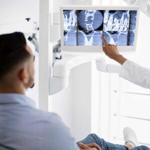
During your initial visit, our oral surgeon will thoroughly review your medical history and discuss any specific concerns you have about your oral health. We’ll perform a comprehensive examination and explain why 3D imaging is recommended for your particular case. You’ll learn how this advanced technology helps us provide safer, more precise surgical care.
We’ll walk you through sample 3D images similar to what we’ll capture of your oral structures, helping you understand exactly what we’re looking for and how the scans influence your surgical plan. Our team will also discuss timing, costs, and insurance coverage for both the imaging and your planned procedure.
Exam Process
What to Expect During the Imaging Procedure
The 3D imaging process is quick, comfortable, and completely painless. You’ll sit in an open scanning chair while our imaging system rotates once around your head, capturing hundreds of detailed images in just 14 seconds. Our experienced technologist will ensure you’re properly positioned and comfortable throughout the brief scan.
There’s no special preparation needed, and you can breathe normally during the scan. Unlike traditional medical CT scanners, our dental imaging system is open and non-confining. The entire process, including positioning and image capture, typically takes less than 5 minutes to complete.
Post-Exam Information
What Happens After 3D Imaging?
There is no recovery period required after 3D dental imaging – you can immediately resume all normal activities, including eating, drinking, and returning to work or school. Our oral surgeon will analyze your scans immediately, often discussing the findings with you during the same visit. We’ll use these detailed images to show you exactly what we’ve found and explain our recommended surgical approach.
Using your 3D scans, we’ll create a comprehensive surgical plan tailored to your specific anatomy and needs. The detailed images become part of your permanent medical record, allowing us to track changes over time and share important information with your other healthcare providers when needed.
We’ll provide you with a clear treatment timeline and detailed pre-surgical instructions based on your specific procedure. Our surgical coordinator will help you schedule your procedure and answer any questions about insurance coverage or financing options. Throughout the entire process, we remain committed to ensuring you feel informed, comfortable, and confident about your upcoming oral surgery.